

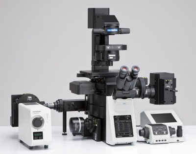
Motorized inverted confocal microscope IX83 (Olympus)
The fully-motorized and automated confocal IX83 microscope represents the most advanced member of the new IX3 series of inverted imaging systems for life science researchers. Microscope provide the ability to perform a multitude of imaging techniques, ranging from long term time-lapse imaging and other demanding cutting edge techniques to casual documentation.
Parameters
- Olympus Disk Spinning Unit (DSU)
- One-deck system
- Lumencor SOLA SEII LED light engine
- ORCA-FLASH 4.0 sCMOS camera
- Metamorph Advanced imaging software
- Advanced Imaging workstation
- Manual and Piezo Z stage
- Kinetics Labmate anti-vibration table
- Operators manual can be found here
MFP-3D-BIO AFM (Asylum Research)
The MFP-3D-BIO™ integrates the atomic force microscope with an inverted optical microscope Nikon TE2000-U. By combining the two technologies, optical data (e.g. functional labeling) can be fused with both quantitative force and topography data. The sample stage of the commercial microscope is replaced with a more rigid mechanical stage called the MFP-3D-BIO™ baseplate, which allows for sub-micron positioning of the AFM tip and sample relative to the optical field of view. The MFP-3D-BIO™ includes TopViewIO functionality, a camera that enables simple alignment of the Superluminescent Diode (SLD) spot on the cantilever and allows positioning of the cantilever over a specific feature on the sample.
Parameters
- AFM performance integrated with optical microscopy
- Simple, high-resolution imaging in liquid on soft biological samples
- Optical navigation of AFM tip for imaging, force spectroscopy and force mapping
- Sub-picoNewton force sensitivity
- Cantilever calibration and automatic elastic Hertz modelling integrated into the AFM software
- 24/7 support from bio-AFM experts (Asylum Research)
- Standard Acoustic Isolation BCH-45
- Operators manual can be found here
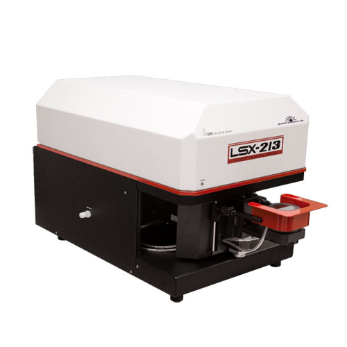
Laser ablation system - LSX-213 G2+ (Teledyne CETAC)
The laser ablation system generates particulate aerosols from solid material by an extremely rapid interaction between high energy UV laser pulses and the sample surface. This process is referred to as ablation. Adjusting laser energy, spot size and pulse frequency using the DigiLaz G2 software optimizes parameters of created pattern (depth, width and profile).
Parameters
- Frequency quintupled, Q-switched Nd:YAG laser, 213 nm
- Spot size range: 4–200 µm
- > 3 mJ/pulse laser energy, computer controlled
- Laser output energy is adjustable from 0–100%
- Flat-top laser beam energy profile
- Laser pulse width: < 5 nsec (typical)
- Laser repetition rate: 1–20 Hz
- Step resolution: 0.16 µm X-Y axes, 0.78 µm Z axis
- Operators manual can be found here
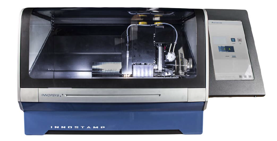
Microcontact printer - INNOSTAMP 40 (INNOPSYS)
Combining microcontact printing flexibility and high-precision automation, the InnoStamp 40 is the perfect tool for nano and micropatterning. A user-friendly system, the InnoStamp 40 allows users to control their processing in order to transfer a wide range of components homogeneously onto defined surfaces. The InnoStamp 40, allows for full automation of nanopatterning processes.
Parameters
- Support for 4 microscope slides and wafer chips with 1 circular stamp (max 96 mm)
- Compatible with micro plates of 96, 384, and 1536 wells and other
- Manipulation of the stamp by magnetic force: micro positioning, high reproducibility & stamp design versatility
- Patterning on non-planar surface, thanks to the use of magnetic force and the flexibility of the stamp (homogeneous pressure)
- Precision: X: +/- 3µm, Y: +/- 3µm, angular: +/- 0.5°
- Compatible inks: nano-objects, chemical substances, biomolecules (proteins, DNA...)
- Microcontact printing applications: for more information about microprinting technology
- Operators manual can be found here
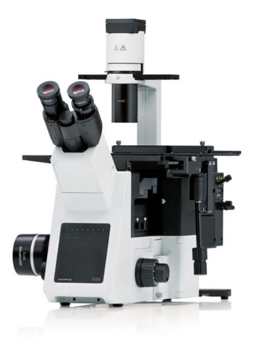
IX53 inverted microscope (Olympus)
The IX53 has been designed from the ground up to be the finest microscope available for routine inverted microscopic analysis. The combination of excellent optical performance and mechanical quality results in a microscope system of outstanding value and comfort for regular use. Features such as the pre-centered phase contrast, relief contrast and the flexible UIS2 DIC optics enable easy visualization across all magnifications for both thin and thick specimens. Available condensers with different working distances and long working objectives allow observation and documentation of even the most complex samples.
Parameters
- Bright-field, phase contrast and fluorescence options are available
- A long working distance condenser
- XM10 monochrome camera 1.4 MP
- Software cellSENS ver. 1.13
- Operators manual can be found here